



Understanding Keratoconus Symptoms and How Eye Care Equipment Detects Them
Keratoconus causes progressive corneal thinning and steepening, so you’ll notice vision that won’t sharpen with new glasses, wavy or ghosted lines, halos and starbursts at night, and increasing light sensitivity. You’re at higher risk with a family history, eye rubbing, asthma, hay fever, Down syndrome, or Ehlers-Danlos. Diagnosis uses slit-lamp exam, retinoscopy, keratometry, corneal topography, and tomography (Scheimpflug or OCT) to map curvature and detect ectasia early. Keep going with Seven OES to see how doctors confirm it and what equipment matters.
Key Takeaways
- Keratoconus causes progressive corneal thinning and steepening, leading to blurry, distorted vision that often doesn’t improve with new glasses.
- Early symptoms include wavy or ghosted lines, halos and starbursts at night, and increased light sensitivity with frequent squinting.
- Night glare worsens over time, and unequal prescriptions may cause headaches or balance strain; both eyes are usually affected asymmetrically.
- Risk factors include family history, chronic eye rubbing, and conditions like asthma, hay fever, Down syndrome, and Ehlers-Danlos.
- Diagnosis uses slit-lamp exam, retinoscopy, keratometry, corneal topography, and tomography (Scheimpflug or OCT); tear film analysis assesses inflammation.
What Are Keratoconus Symptoms and Who Is at Risk?
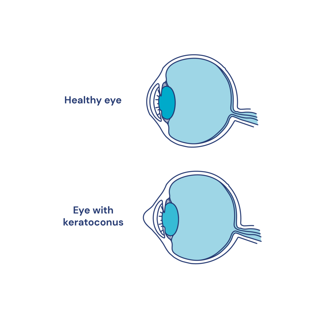
Keratoconus is a progressive thinning and steepening of your cornea that makes it bulge into a cone shape, causing blurry, distorted vision and light sensitivity.
You’ll notice keratoconus early symptoms as vision that won’t sharpen with glasses, glare that worsens at night, and frequent prescription changes. It usually involves both eyes but can be worse in one.
Who’s at risk? You’re if you have a family history, rub your eyes vigorously, or live with asthma, hay fever, Down syndrome, or Ehlers-Danlos. It often starts in your late teens to 30s and may progress for a decade or longer.
Ask early: Are night halos or sudden clouding new? If yes, seek evaluation with keratoconus diagnosis equipment such as corneal topography and tomography to document progression.
Early Keratoconus Symptoms Patients Should Watch For
Noticing vision that won’t fully clear with new glasses or contacts? That’s a common early clue.
Do straight lines look wavy or ghosted? Do you see double in one eye, especially when the other is covered? Are halos or starbursts around headlights new or worsening? Do lights feel uncomfortably bright? Do you squint more, yet still feel blur that shifts day to day? Is one eye clearly worse, with frequent prescription changes?
Watch for headaches or balance strain from unequal prescriptions. Track increased nearsightedness and irregular astigmatism that didn’t bother you before. Avoid eye rubbing; it can aggravate changes.
If these signs ring true, ask for keratoconus detection tools, including a corneal topographer for keratoconus.
Early confirmation guides timely monitoring and protects long-term vision.
How Ophthalmologists Diagnose Keratoconus Accurately
Although symptoms raise suspicion, ophthalmologists confirm keratoconus with targeted tests that map and measure your cornea.
You’ll start with an extensive eye exam focused on your blurry vision, glare, and distortion. Expect a refractive assessment using a retinoscope and phoropter to reveal irregular astigmatism patterns that don’t correct cleanly.
Next, a slit-lamp exam inspects corneal shape and clarity under magnification. Corneal topography captures detailed maps, highlighting early cone formation and progression over time.
Keratometry then checks curvature by analyzing reflected rings; steep, asymmetric readings suggest ectasia.
These steps use precise ophthalmic equipment for eye diseases to separate keratoconus from other causes of visual strain and headaches.
The result: an evidence-based diagnosis that guides timely monitoring and customized vision correction without delay.
Ophthalmic Equipment Commonly Used to Detect Keratoconus
From clinic to corneal map, precise instruments pinpoint early keratoconus before vision rapidly worsens.
Which tools matter most when your glasses stop working and astigmatism keeps changing? Start with slit-lamp biomicroscopy to spot corneal thinning, scarring, and Vogt striae. Use retinoscopy to detect the scissor reflex that flags irregular optics.
Need objective shape data? Corneal topography measures anterior curvature to reveal asymmetric steepening. Tomography (Scheimpflug or OCT) adds posterior elevation, pachymetric maps, and Belin/Ambrósio indices—critical for subclinical disease.
Ultrasound pachymetry confirms minimum corneal thickness when imaging is limited. Placido-based systems track progression; compare K-values and cone location over time.
Concerned about surface quality? Tear film analysis can show raised IL‑6 and TNF‑α, supporting inflammatory activity.
Together, these devices detect, stage, and monitor progression early.
Where Clinics Can Find Reliable Ophthalmic Equipment for Keratoconus Testing
When your keratoconus patients progress faster than lenses can compensate, you need vetted sources for dependable devices—now.
Where should you look? Start with Seven OES ophthalmic division: 15+ years in South America, now serving North America, built on efficiency, security, and profitability.
Need multi-brand options? We’ve specialized since 2010, scaling to Latin America’s largest used equipment maintenance and sales provider by 2021, and launched U.S. operations in 2022.
Ask: Are units reviewed, calibrated, and certified? Yes—every device undergoes thorough maintenance and ships with traceable Calibration Certificates.
Want diagnostic confidence? Expect innovative, high‑quality systems, compliant service, and a 1‑year warranty.
Prefer efficient workflows and affordability? They prioritize user‑friendly solutions.
Next steps: book a FREE one‑on‑one consultation, call or email support, browse the online shop, and sign up for first‑time discounts.
Frequently Asked Questions
Can Keratoconus Progression Be Slowed With Lifestyle Changes?
Yes. You can likely slow progression by avoiding eye rubbing, treating allergies aggressively, using preservative-free tears, wearing UV-blocking sunglasses, managing screen strain, sleeping on your back, and keeping follow-ups. Ask about corneal cross-linking early if changes appear.
Are Contact Lenses or Glasses Better for Early Keratoconus?
Glasses may work initially, but contact lenses usually give sharper vision earlier. You’ll likely benefit from soft toric or early hybrid/scleral lenses. Monitor glare, ghosting, eye rubbing. Ask about cross-linking, allergy control, and regular topography to track changes.
How Often Should Keratoconus Patients Schedule Follow-Up Exams?
Schedule follow-ups every 3–6 months; go sooner if vision blurs, halos increase, night glare worsens, or lenses feel uncomfortable. After corneal cross-linking, visit at 1 week, 1–3 months, then 6–12 months. Ask about topography frequency.
What Insurance Considerations Apply to Keratoconus Treatments and Testing?
You’ll check if your plan covers cross-linking, specialty contacts, topography, and medically necessary lenses. Verify prior authorization, deductibles, CPT/ICD codes, in-network providers, and out-of-pocket caps. Ask about replacement frequency, fitting fees, denials, and appeals.
Can Keratoconus Affect Eligibility for Refractive Surgery Later?
Yes. You’re typically not eligible for LASIK/PRK with keratoconus due to corneal instability. Have you had topography, thickness, or progression checks? Consider cross-linking, specialty lenses, or staged options. Ask about risk, stability criteria, and alternatives like ICLs.
Conclusion
You’ve now got a clear depiction of keratoconus—who’s at risk, what early symptoms look like, and how ophthalmologists confirm it with precise imaging. Noticing night glare, halos, frequent prescription changes, or eye rubbing? Don’t wait. Ask for corneal topography, tomography, pachymetry, and aberrometry. Wondering which devices are reliable? Compare validated platforms, check service support, and verify calibration standards. Ready to act? Book a screening, track symptoms, and partner with a clinic that invests in quality ophthalmic equipment.


